Meniscal Fibrocartilage of the Knee: Review of Current Evidence
Christopher Hope, SPT
Background
Last year I damaged the meniscus in my right knee when I slipped on ice in a parking lot. After six months of symptom management and reduced activity, I elected to proceed with surgery in hopes of returning to my prior level of activity. Considering the extent and location of damage, a partial meniscectomy was performed instead of the originally intended meniscal repair. I was very disappointed in that decision because I knew my risk for developing osteoarthritis had just increased significantly. This experience encouraged my interest in the menisci of the knee.
During my physical therapy rehabilitation, I was also enrolled in two courses that would ultimately contribute to this project. The first, Evidence Based Practice II, required the completion of both a critically appraised paper and critically appraised topic. Considering my ongoing personal experience with meniscal injury, surgery, and rehab, I chose for my topic: effects of strength training on knee osteoarthritis following partial meniscectomy. For the second course, Advanced Orthopaedic Assessment and Treatment, my final paper was titled Review of Meniscal Fibrocartilage in the Knee. Resources and knowledge acquired from both of these assignments contributed to this project.
Overview & Purpose
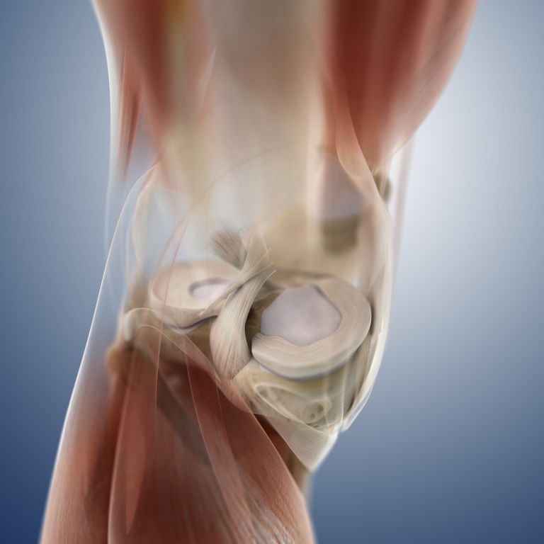
Meniscal pathologies are related to its function and role, which in turn are directly related to its properties and characteristics. Cellular and extra-cellular components (i.e. collagen fibers) influence the ability of the meniscus to resist stress, load-bear, and absorb shock. The location, shape, and attachments of the menisci further influence its function and role.1,2
Primary roles of the meniscus in the knee include load bearing and increasing the contact area between the femur and the tibia, and subsequently decreasing contact pressure and stress on articular cartilage. Other functions of the meniscus include shock absorption and assisting in knee joint stability.1,2
Understanding the role of the meniscus during arthrokinematic knee motion is important to understanding injury mechanisms. For example, it helps to explain why compression with rotation is a common mechanism of meniscal injury.3 There are several types of meniscal tears, generally grouped as either degenerative or traumatic.1 Healing is largely dependent upon blood supply, and since only the outer one-third of the meniscus is vascularized, this portion tends to heal well while the central portion has a significantly reduced ability to self-heal.2
A number of tools are available to aide clinicians in diagnosing a meniscal tear.4 Reviewing the current literature will help to identify which of those tools has the most evidential support, and how to best use the tools at a clinician’s disposal. Finally, there are also a number of interventions available to address meniscal injury, generally grouped as either surgical or non-surgical.3 Again, reviewing the current literature will provide insight as to the efficacy of these interventions and subsequent rehabilitation.
The purpose of the presentation is to: first, briefly review the properties, characteristics, function, and role of the meniscus and their collective influence on injury and healing; and second, review the current literature regarding diagnostic tests and intervention and treatment.
Products
An audiovisual presentation has been created to fulfill the purpose listed above. The presentation is intended as a review of current evidence related to meniscal fibrocartilage of the knee for practicing clinicians who treat meniscal injuries or conduct post-surgical rehabilitation. The presentation is made available through the VoiceThread application.
The presentation will be made available to physical therapists in an outpatient orthopedic setting that has a strong emphasis on sports injuries and related orthopedic surgeries (Pivot Physical Therapy). Discussion with the clinic director (and committee member, Chris Kosobucki) confirmed that information pertaining to meniscal fibrocartilage of the knee would be relevant and helpful to the clinicians of this practice.
Also, an educational brochure has been developed to review information related to the meniscus and meniscal surgery. The brochure is intended for patients who present to a physical therapy clinic (Pivot Physical Therapy) with meniscal pathology and includes information on the meniscus, types of surgery, factors considered for surgery, and benefits of physical therapy.
Evaluation
Feedback from those who review the presentation will be collected through a link to a custom survey through SurveyMonkey. The address is provided within the presentation, but can also be accessed using the link above. There are seven questions that are answered using a ratings scale of 1 to 5, with 5 being the most favorable score on each question. A few practicing physical therapists, students, and other healthcare professionals were willing to provide initial feedback in order to determine the initial quality and effectiveness of the presentation. The lowest score received was 4/5, with the overall average score being 4.87/5 points. Comments were mostly complementary, with some providing critical evaluation (as requested) and suggestions for improvement.
To obtain initial feedback, the brochure was provided to a few patients currently receiving physical therapy for meniscal injury. The intention of the feedback form was to judge the literacy level, appearance, and effectiveness of the brochure. There are 6 questions that are answered using a ratings scale of 1 to 10, with 10 being the most favorable score on each question. Collected scores varied greatly, ranging from 2/10 (“I already had surgery”) to 10/10. The scores and comments were generally positive, with the overall average score being 8.16/10 points.
Acknowledgements
The first person who needs to be acknowledged is my wife, Jessica, who has a full appreciation for the demands and stress of this level of academic endeavor. Through it all, she has been the epitome of grace, support, and encouragement. Thank you sincerely for making it possible for me to pursue this dream.
Second, thank you to the UNC DPT faculty for investing in my education and my career. Special thanks to Jon Hacke for not only being my advisor and role model, but for making this all possible in the first place. You changed my life. Thank you. Also special thanks to Mike McMorris, another role model, for taking the time to illustrate what it looks like to invest in someone else. I appreciate your time and the clinical (and life) lessons you gave me. Finally, a special thanks to my capstone advisor Mike Gross, the Jim Morrison of orthopedics: brilliant, influential, and iconic. Thanks for sharing your stories, wisdom, and knowledge.
Third, and finally, thank you to those coworkers from days past who facilitated my progress towards where I am today and showed me what it looks like to be where I want to be. This includes my capstone committee members Chris Kosobucki and Gary Johnson, along with so many others who have directly and positively influenced my path. Thank you for building and solidifying my faith in this truly special profession.
References
- Arnoczky et al: Chapter 12 Meniscus. In Woo SL, Buckwalter JA (eds): Injury and Repair of the Musculoskeletal Soft Tissues. Park Ridge, Illinois, American Academy of Orthopaedic Surgeons, 1988, pp 487-537
- Hartigan E, Lewek M, Snyder-Mackler L. Chapter 11 The Knee. In Levangie PK, Norkin CC: Joint Structure and Function. 5e. Daryaganj, New Delhi, India. Jaypee Brothers Medical Publishers (P) Ltd; 2012, pp 395-439
- Hertling D, Kessler RM. Chapter 15 Knee. In Hertling D, Kessler RM: Management of Common Musculoskeletal Disorders. 4e. Philadelphia, PA. Lippincott Williams & Wilkins. 2006; pp 487-558
- Magee DJ. Chapter 12 Knee. In Magee DJ: Orthopedic Physical Assessment. 6e. St. Louis, Missouri. Elsevier Saunders. 2014; pp 765-887
- Picture: https://fthmb.tqn.com/siP6tf9k4Ask5hsQUe6-ornqNDo=/768×0/filters:no_upscale()/about/168835293-56a6d9833df78cf772908bd2.jpg
4 Responses to “Meniscal Fibrocartilage of the Knee – Review of Current Evidence”
Christopher Hope
Hi Christie! Thanks for taking the time to review my capstone material and provide feedback. I appreciate your compliments and I’m glad you’ll be able to use some of the resources.
The comment from Howell, et al regarding extensor strength is comparing quad strength between operative and non-operative extremities. The authors warn that even with diligent rehabilitation, recovery of pre-surgical extensor strength may take 4-6 weeks following arthroscopic partial meniscectomy. Even then, there might remain a discrepancy in quad strength between operative and non-operative extremities that increases a patient’s risk of injury on return to sporting activity. This is where the guidelines for 80% and 90% are suggested for return to practice and return to game.
Regarding the measurement of knee extensor strength, please see my comments in response to Katie’s similar question above. Bottom line is that I have not come across evidence that suggests any one method is a better predictor of success upon return to sport, and I anticipate that a combination of strength assessments will produce a more accurate prognosis. I agree that testing procedures would need to be clearly communicated and consistent in order to promote accuracy. If you come across any research that helps address this question, please share.
Again, thanks for your feedback and for the opportunity to clarify the research.
Christie Clem
Hope,
Nice work on your capstone! I like the personal touches that you added to your VoiceThread and the Capstone website by including your wife and son. Anyone that spends time with you knows how important they are in your life and that is easy to see in this project as well.
The patient handout is a great resource. It’s visually appealing and easy to read. So many times I would have liked to have some kind of educational material to give a patient, but there never seems to be enough time to create one. This will be a handout that I add to my collection to have on-hand when it is needed.
The VoiceThread had clearly defined learning objectives in addition to the information being presented in a very logical order. I appreciate the large number of evidence that was considered when identifying the best diagnostic tests for meniscal tears along with the inclusion of the Osteras et al and Noyes et al articles for interventions following surgery. I plan to look up these articles and add them to my personal collection for future reference.
The information from Howell et al stated the need for 80% extensor strength to return to practice and 90% extensor strength to return to games. Was this strength percentage comparing the current strength to that of the non-operative leg or to the pre-surgical strength measurement on the side that had surgery? My first thought was the non-operative side, but a bullet point on that slide mentions the importance of returning to pre-operative strength. Also, was there any mention of how the strength measurements were taken…isokinetic testing, a dynamometer, MMT? Of these I would think dynamometry would offer the most objective and feasible combination in most clinics. But like any other form of measurement, testing procedures would need to be clearly defined and performed in a consistent manner to get accurate results.
I think you created a very thorough capstone can be used to help both practitioners and patients. Great job!
Christie
Christopher Hope
Hi Katie! Thanks for taking the time to review my material and offer feedback. Your survey comments are very helpful, and I appreciate your insight.
Thanks for asking about using a cluster of tests for diagnosing meniscal pathology. To answer your first question: no, I would not recommend always referring a patient for MRI if you suspect a tear. The issue of unjustified arthroscopies and overuse of diagnostic imaging technology is well established. Additionally, arthroscopic examination has been used to confirm that MRI has about the same diagnostic accuracy (80%) as Apley’s test (80%) and McMurray’s test (79%).(1) Moreover, MRI has been shown to have a false positive rate of 65% for the medial meniscus and 43% for the lateral meniscus.(2) An MRI is obviously a helpful tool with useful diagnostic benefits, but it should be considered for use to rule out a meniscal tear only after careful physical therapy evaluation and examination.
To answer your second question: yes, there is evidence to show that a combination, or cluster, of special tests can be very effective in predicting meniscal tears. The cluster consists of five tests: patient report of catching or locking, pain with forced hyperextension, pain with maximum flexion, pain or an audible click with McMurray’s maneuver, and joint line tenderness to palpation. A positive finding on all five of these tests has been found to have a 92% positive predictive value for identifying a meniscal tear. A positive finding on three or four tests still produced a positive predictive value >75%. Concurrent ACL pathology and/or DJD alters these number, so a clinician should not rely on physical examination findings alone when it’s suspected that additional structures are injured.(3)
Finally, while it is important to recover knee extensor strength following meniscal injury, I have not come across evidence that suggests any one method is better than another in measuring such strength. There are many published studies documenting the prevalence and severity of decreased quad strength following knee injury, including at least a few systematic reviews, but I don’t see where research has been conducted to identify the best, or most effective/accurate, method of measuring quad strength following injury or surgery – especially in regards to predicting a patient’s readiness for return to sport. As with diagnostic tests, my inclination is that a combination of tests (i.e. functional, isokinetic, dynametric, manual) would best indicate a patient’s readiness to return to his/her prior level of activity.
Again, thanks for your review and for the opportunity to expand on these topics.
Resources
1.Rinonapoli G, Carraro A, Delcogliano A. The clinical diagnosis of meniscal tear is not easy. Reliability of two clinical meniscal tests and magnetic resonance imaging [abstract]. Int J Immunopathol Pharmacol. 2011. 24(1 Suppl 2): 39-44
2.Ben-Galim P, Steinberg EL, Amir H, Ash N, Dekel S, Arbel R. Accuracy of magnetic resonance imaging of the knee and unjustified surgery. Clin Orthop Relat Res. 2006. 447: 100-4
3.Lowery DJ, Farley TD, Wing DW, Sterett WI, Steadman JR. A clinical composite score accurately detects meniscal pathology. Arthroscopy. 2006. 22(11): 1174-9
Katie Sly
Hi Chris!
I enjoyed reviewing your materials. I especially loved seeing the picture of you and your son at the beginning of your Voice Thread. I’m glad to see him in Carolina blue!
I wanted to inform you that I filled out your survey on Survey Monkey. Also, I wanted to let you know that your CAT is not linked correctly, so you might want to look into that.
Now for my comments:
-I thought your review of the anatomy and functions of the meniscus was comprehensive and informative. Additionally, I thought you spoke clearly in your presentation and met your objectives.
-I thought your brochure was visually appealing and provided good information to patients about different types of meniscus surgeries. From my clinical rotations, I know many patients benefit from further education about this topic.
-Your literature review was very informative. Since multiple special tests do not have good accuracy, would you recommend always referring a patient to receive an MRI if you suspect a tear? Is there any evidence to show that a combination of positive special tests is as accurate as an MRI?
-I was also interested to learn that gaining extensor strength is especially important in return to sport. You cited that 80% of strength should return before practice and 90% before game. Would you recommend using isokinetic testing or some form of dynamometry to measure strength in this case or would MMT be sufficient?
Again, great job, Chris! I really enjoyed reviewing your capstone! I can tell a lot of hard work went into your products!
Thanks,
Katie Sly